Velocitas VMI Applications
The Velocitas VMI Spectrometer is used in Velocity Map Imaging experiments with laser pulse durations from nanoseconds to attoseconds and photon wavelengths from IR to XUV and X-ray. Capturing 3D information on kinetic energy, angular distribution and signal intensity, the spectrometer can be used as a diagnostic tool in femtosecond and attosecond laser characterisation, or in fundamental investigations of electron processes, stereochemistry and photodissociation dynamics.
Specific examples of Velocitas VMI applications include:
Photoelectron Spectroscopy
Photoelectron spectroscopy provides fundamental information on electronic structure in atoms and small molecules, playing an integral part in investigations into electron dynamics and coherent control.
Attosecond Laser Characterisation
By observing the intensity and angular distribution of photoelectron interference patterns from XUV-IR photoionisation, the relative phase is known and the electric field of attosecond pulses can be reconstructed.
Photodissociation Dynamics
Imaging the 3D velocity distribution of state-selected reaction products helps build a picture of molecular stereodynamics and how it affects chemical reactions and energy transfer processes.
Time Resolved Dynamics
Fundamental dynamical processes can be characterised by recording photoelectron and/or photoion kinetic energy distributions for different delays in pump-probe spectroscopy experiments.
VMI is an increasingly popular technique in a wide range of research laboratories. We aim to understand your specific application to be able to offer a bespoke spectrometer design to fit your individual needs.
Contact us to discuss your needs >
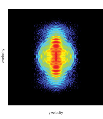
Above: VMI of photoelectrons from Above Threshold Ionisation (ATI) of Xe in VMI PRIME.
The image shows the raw, 2D photoelectron angular distribution from strong field ionisation of xenon, recorded in VMI PRIME with 800 nm laser pulses of duration 35 fs and intensity 1 x 10¹⁴ Wcm-2. The imaged electrons lie on concentric rings, with each successive ring indicating the absorption of another photon, consistent with above threshold ionisation. The presence of side lobes is a result of interference effects, where an emitted electron will rescatter from the atomic core with characteristics representative of the carrier envelope phase (CEP) of the ionising laser pulse.
With permission from MBI, Berlin.
Velocitas VMI systems have been used in internationally renowned research institutions, including:
Max Born Institut, Berlin, in partnership with Prof Marc Vrakking, to investigate above threshold ionisation and time resolved IR-XUV spectroscopy of atoms.
University of Bristol, UK, in partnership with Prof Mike Ashfold to investigate resonant one-colour and two-colour photoionisation of diatomic molecules, with dc slicing for efficient data analysis.
Sandia National Laboratories, California, in partnership with Dr David Chandler, to investigate photodissociation properties of small molecules.